|
|
|
|
|
|
Kære DOS-medlem
DOS Kongressen 2025 finder sted onsdag – fredag i uge 46, dvs. 12.-14. november 2025 ved Scandic Falkoner.
|
|
Sæt kryds i kalenderen og vi sender yderligere info, når hotelbooking med rabat og tilmelding åbner til sommer.
Abstract submission er nu åben og du kan indsende dit bidrag frem til 15. april.
Vi skal også have fundet prisvinderne for årets bedste publicerede artikel og forsvarede ph.d. Deadline: 1. april 2025.
|
|
Danish Orthopedic Academy (DOA) er kommet godt fra start og har fået sin egen hjemmeside. Den opbygges løbende og vil indeholde vigtige info - men bare rolig, som DOS medlem får du også tilsendt info som nyhedsbrev. https://doacademy.dk/
|
|
Acta Orthopaedica har igennem 2024 haft et social media udvalg. Arbejdet bliver yderligere professionaliseret i den kommende tid, nu hvor vores kollega Aleksi Reito fra Finland er blevet ansat som Social Media Editor. Se nærmere i nedenstående nyhedsbrev fra Acta, som du også kan abonnere separat.
|
|
Young Nordic Orthopaedic Federation Sidst men ikke mindst, vil vi opfordre alle uddannelsessøgende læger til at deltage i denne spørgeskema-undersøgelse fra NOF vedr. yngre lægers selvstændighed.
De bedste hilsner
|
|
Jan Duedal Rölfing
DOS editor
|
|
Please find below highlights of recently published articles.
Acta SoMe co-editor
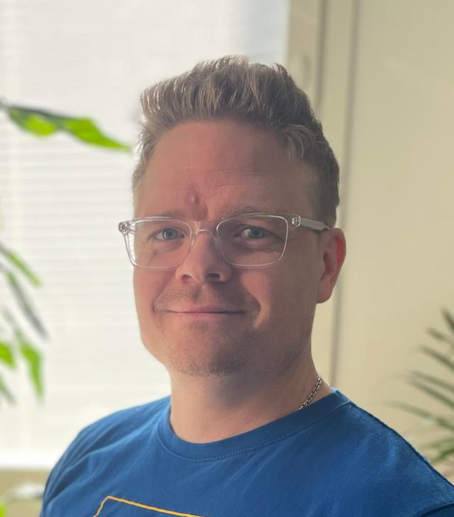
I am pleased to announce that the first social media (SoMe) co-editor of Acta has been appointed.
|
|
|
Associate professor, MD, PhD, Aleksi Reito, has joined the editorial team.
He has been active on SoMe for a long time and is a member of the SoMe group of Acta. He is an experienced orthopedic surgeon and researcher.
You may already have discovered the nice infographics of the articles on LinkedIn
With his help, Acta will be more active on social media and get more visibility. Please follow us.
|
|
|
In 2024, all articles are available as one issue in volume 95 (2024). They are categorized within subspecialties and are available on our website.
|
|
|
On behalf of the Editorial team,
|
Søren Overgaard
Editor-in-chief
|
|
|
|
Join our mailing list
|
|
|
This newsletter may have been forwarded to you as a NOF member by your national society head (Denmark, Estonia, Finland, Iceland, Lithuania, Norway, Sweden, the Netherlands). This mode of distribution can, however, be slow or unreliable due to outdated contact information. An alternate way to ensure that you receive this newsletter directly as it is published is to join our mailing list by clicking on the link below.
|
|
|
|
|
|
|
|
|
|
|
Follow us on social media to read newly publishes articles
|
|
|
|
|
|
|
|
|
|
|
|
Selected Highlights
|
|
|
|
|
|
|
In the field of orthopedic diagnostics, the integration of artificial intelligence (AI) is an emerging topic, aiming to enhance accuracy and efficiency in fracture detection. The recent study by Axenhus et al., titled "Automated diagnosis and classification of metacarpal and phalangeal fractures using a convolutional neural network: a retrospective data analysis study," marks a significant advancement in this domain.
Hand fractures, particularly those involving the metacarpal and phalangeal bones, are prevalent injuries encountered in emergency departments. Despite their frequency, diagnostic errors remain a concern, potentially leading to complications and delayed treatments.
Axenhus and colleagues addressed this challenge by developing a convolutional neural network (CNN) model designed to diagnose and classify these fractures using plain radiographs. Their model achieved a mean weighted area under the curve (AUC) of 0.84, with sensitivity and specificity rates of 86% and 76%, respectively. Notably, the model excelled in identifying transverse metacarpal fractures (AUC = 0.91) and tuft phalangeal fractures (AUC = 0.97), demonstrating 100% sensitivity in both categories. These findings underscore the potential of AI-driven tools to augment clinical decision-making in orthopedic practice. Accurately identifying and classifying hand fractures, such models can serve as valuable adjuncts to clinicians, potentially reducing diagnostic errors and improving patient outcomes, but also reducing work-load and the possibility to shift attention to other tasks.
However, the integration of AI into clinical workflows necessitates careful consideration. While the performance metrics are promising, the model's efficacy in diverse clinical settings and its adaptability to varying imaging protocols require further validation. Moreover, the ethical implications of AI in healthcare, including issues of transparency and accountability, must be addressed to foster trust among practitioners and patients alike.
In conclusion, the study by Axenhus et al. represents a step toward the use of AI in orthopedic diagnostics. As technology continues to evolve, collaborative efforts between clinicians and data scientists will be essential to harness the full potential of AI, ultimately enhancing the quality of patient care in orthopedics
Co-editor
Taco Gosens
|
|
|
|
Vascularized fibular grafting following tumor resection demonstrates acceptable long-term outcomes in Denmark: a national retrospective cohort study
Christian Lind Nielsen, Daniel Thor Halberg Dybdal, Peter Vester-Glowinski, Lisa Lyngsie Hjalgrim, Pernille Edslev Wendtland, Birgitte Jul Kiil, Michael Melchior Bendtsen, Michael Mørk Petersen, Thomas Baad-Hansen
Acta Orthopaedica, 96, 2025, 87–93.
|
|
Bone sarcomas, such as osteosarcoma and Ewing sarcoma, are rare primary malignant bone tumors requiring wide or marginal resection for survival. Vascularized fibular grafting (VFG) represents an often used biological reconstruction in orthopedic oncology. Its benefits include size, and shape, and when used as a vascular graft blood supply promoting bony healing via hypertrophy. VFG is typically applied for diaphyseal reconstruction alone or in combination with allograft and for intraarticular reconstruction in the upper extremity. Despite its advantages, there remains a relatively high rate of postoperative complications including delayed union and graft fracture, which may require revision surgery.
Nielsen et al. present a retrospective evaluation of a national Danish cohort comprising 27 patients undergoing biologic reconstruction with VFG. The indications were 13 cases of Ewing sarcoma, 12 cases of osteosarcoma, and 2 cases of giant cell tumor. The median age at surgery was 16 years with median follow-up of 82 months. Osteosynthesis was achieved through internal fixation in all cases. The overall rate of graft union was 67%, with a median time to union of 13 months. 20 patients experienced complications necessitating surgical intervention. 2 patients in the cohort underwent above-the-knee amputations during the follow-up period representing a limb salvage rate of 93%. A transient postoperative peroneal nerve palsy occurred in 7 patients, with all cases resolving spontaneously. The 5-year overall survival rate was 81%. Patients with upper extremity tumors were more likely to attain graft union and were less likely to undergo multiple revisions than patients with lower extremity tumors.
Biologic reconstruction using VFG remains a useful option to reconstruct diaphyseal bony defects in adolescents undergoing surgery for malignant bony tumors even with the development of custom-made endoprostheses. The risk of revision surgery remains relatively high and this risk is smaller in patients undergoing upper extremity (humerus) reconstruction.
Co-editor
Ilkka Helenius
|
|
|
|
|
|
|
|
|
|
|
|
|
|
 |
|
|
|
|
|
|
|
|
|
|